Positive Health Online
Your Country

![[Image: http://www.abundanceandhealth.co.uk]](/img/original/BannerAvatar/2836.df7d2315253110ff5b3a566f94871f99.jpg "http://www.abundanceandhealth.co.uk")
![[Image: http://www.drsgoodman.com/books-goodman/52-nutrition-and-cancer]](/img/original/BannerAvatar/1859.1296090fd8385e7ceff51c8011559097.jpg "http://www.drsgoodman.com/books-goodman/52-nutrition-and-cancer")
![[Image: Lotus Books]](/img/original/TopicbannerAvatar/953.adb373b0210368e0c9ce4d9a8a9f01aa.jpg "Lotus Books")
![[Image: Turning Point]](/img/original/TopicbannerAvatar/981.28945ea95f14bc185effb4ae6d8a75ab.jpg "Turning Point")
Manipulation to the Foot (Medial Border)
listed in osteopathy, originally published in issue 210 - November 2013
Introduction
The operator should insure that they have a good knowledge of the foot’s surface anatomy thereby enabling them to palpate/locate the intrinsic bones of the foot, and their associated joints.
The most important anatomical landmark of the medial border is the navicular tubercle.
![]()
Medial Border
This consists of the following joints
- Talo/navicular;
- Navicular cuneiform;
- Cuneiform metatarsal;
- Metatarsal/interphalangeal.
The key to carrying out successful foot manipulation is to keep one of the bones of the joint fixed whilst moving the other bone, usually the distal one.
It is important to maintain dorsiflexion of the ankle mortice thereby locking it, as well as limiting movement of the talus and calcaneum. Thereby ensuring specific movements of the joints of the medial border of the foot.
Talo-Navicular Joint
Type: Modified Ball and Socket
Technique
- The operator locates the talus with the web of their hand;
- With the lower hand, the index finger is placed around the navicular and brings the foot into dorsiflexion;
- With both arms and elbows locked (this will prevent RSI in the operator), the operator is able to move the navicular in a circumductory movement around the fixed talus by the operator using their body weight to initiate motion, bending their knee where necessary, thus insuring correct posture;
- When dysfunction is encountered a rapid low velocity impulse is applied with minimum depth across the joint.
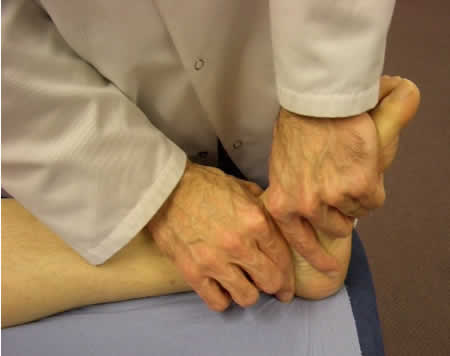
Fig 1: HVT to Talo-Navicular Joint
Navicular-Medial Cuneiform Joint
Type: Multi-faceted Plane
Technique
- The operator wraps the web of their lower hand around the navicular (using the navicular tubercle as a landmark) whilst also maintaining dorsi-flexion;
- The index finger of the lower hand is wrapped around the medial cuneiform;
- With the navicular fixed, again with the operator using fixed elbows, the cuneiform is sheared into dorsi/plantar flexion. A small amount of medial/lateral shearing is also possible;
- When dysfunction is encountered, a rapid low velocity impulse is applied with minimum depth across the joint.
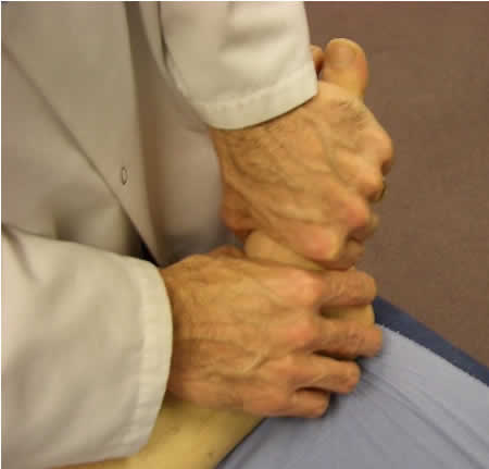
Fig 2: HVT to Navicular-Med Cuneiform Joint
Medial-Cuneiform First Metatarsal Joint
Type: Hinge/Plane Joint
Technique
- The operator wraps the web of their hand around the medial-cuneiform whilst simultaneously maintaining dorsi-flexion;
- The operator then places their thumb over the dorsal aspect of the first met, whilst placing the pad of their index finger under the plantar aspect of the first met, thereby creating a clamp like grip;
- With the medial-cuneiform fixed, the operator is again using a fixed elbow to apply a hinge-like motion through the joint, whilst maintaining correct posture and bending their knee where necessary;
- When dysfunction is encountered a rapid low-velocity impulse is applied with minimum depth across the joint.
N.B. Always re-motion test following manipulation.
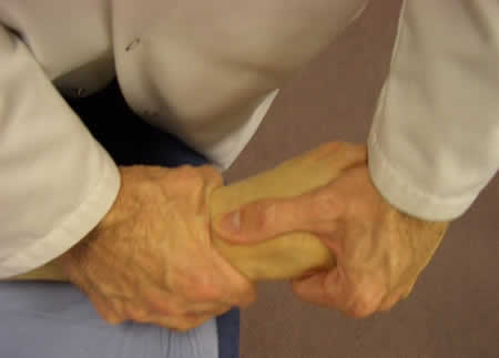
Fig 3: HVT Med Cuneiform-1st Metatarsal Joint
Comments:
-
Professor/Dr Brian A Rothbart said..
The foot manipulations procedures Dr Lintonbon described in his article - Manipulation of the Foot - are indeed effective interventions for repositioning the medial column back into its anatomical neutral position.
However, the issue that needs to be determined is why do the foot subluxations occur. Is it due to trauma, pathology or possibly abnormal inherited foot structures.
If the foot subluxations are the result of trauma to the medial column of the foot, the foot manipulation techniques described by Dr Lintonbon would be my intervention of choice. But if the foot subluxations are the result of either The PreClinical Clubfoot Deformity or Rothbarts Foot (inherited abnormal foot structures) then manipulation of the foot will not premanently resolve the problem.
Professor Rothbart