Positive Health Online
Your Country

![[Image: http://www.abundanceandhealth.co.uk]](/img/original/BannerAvatar/2836.df7d2315253110ff5b3a566f94871f99.jpg "http://www.abundanceandhealth.co.uk")
![[Image: http://www.drsgoodman.com/books-goodman/52-nutrition-and-cancer]](/img/original/BannerAvatar/1859.1296090fd8385e7ceff51c8011559097.jpg "http://www.drsgoodman.com/books-goodman/52-nutrition-and-cancer")
Add as bookmark
Symbionts and their Role in Health and Nutrition
listed in naturopathy, originally published in issue 143 - January 2008
In this article I explain why symbionts are key factors to health and well being of the body. Symbionts are produced when plant food is digested by certain lactobacilli. They are absorbed into the blood and tissues and are indispensable agents for all processes in the body. Certain food fermentation products contain different levels of these indispensable symbionts.
What is a Symbiont?
The term Symbiont (sym= together bios= life) describes microscopic organisms, smaller than cells, that live together with highly developed multicellular organisms, including human beings. Symbionts can easily be seen in live blood cultures using dark field microscopy* at a magnification of 1,000 x. They appear as actively moving particles in the blood plasma (liquid part of the blood), and are approximately 1/20 the size of a red blood cell. Symbionts are the building blocks for all metabolic processes. Their abundant presence in the plasma is a clear sign of a favourable acid-base balance and healthy immune function. Together with minerals and trace elements, they build the fundamentals for optimal energy production and balanced, healthy metabolism. Abundant symbionts prevent viruses, bacteria and parasites from developing into a disease process.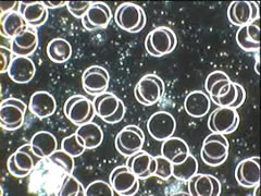
Blood with symbionts as viewed via dark
field microscopy
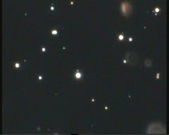
Vita-Biosa
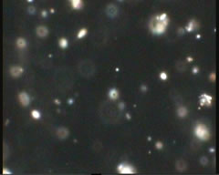
Regulat
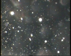
Symlixir
The small white specks represent
symbionts and are in continual vibrational
movement. The background of the
Symlixir sample appears brighter due to
the huge number of symbionts
Dark field microscopy practitioners have several different terms for symbionts: symprotit, spermit, chondrit, etc., according to their shape, movement, characteristics and hypothesized stage of development. Symbionts are believed to be conglomerates of even smaller submicroscopic particles called ‘Protits.’** Chemically, they are believed to consist of mainly albumin and globulin, which are proteins that serve as a buffer against metabolic imbalances. They do not have cell walls, nor a fixed form, and can change their appearance within seconds. They are too small to be bacteria and do not seem to contain larger amounts of DNA or RNA. The most curious aspect is the self-movement of the symbionts. They move in a slow, vibrational, 3-D arbitrary movement that resembles the characteristic of Brownian molecular movement.
The Major Role of Lactobacilli and Bifidus Bacteria
Why do we take probiotic products? Why, exactly, do we believe that the intestinal flora plays such an important role in metabolism and immune function? Is the sole purpose of taking probiotics in the diet to produce lactic acid?I propose to demonstrate that the major role of lactobacilli and bifidus bacteria is to produce symbionts. It is crucial that symbiont production take place at the very beginning of the small intestine under the ‘cleanest’ possible conditions, and in the presence of plentiful nutrients.
Symbionts, Protits and their Struggle with Mainstream Science
Symbionts have not been sufficiently researched by mainstream science and, therefore, have no official definition. Mainstream medicine (that widely ignores the use of the dark field microscope) views symbionts as lifeless globulin particles resulting from the red blood cell breakdown process or as artifacts (meaningless optical disturbances). When one observes a live blood culture under a dark field microscope, it is difficult to imagine these actively moving and interacting symbionts to be ‘dead’ matter.Symbionts can only be observed in live blood cultures using a dark field microscope. The bright-field microscope does not allow the observation of live blood or tissues, because the blood/tissue sample needs to be dried and stained in order to be looked at. This process kills and dissolves all ‘living’ particles, which can then no longer be observed.
Antoine Béchamp: “All cells, organs, all living forms are built from these little bodies.’’
When Anton Leeuwenhoek developed the first microscope in the 17th century these particles that we now call symbionts where already observed. In his famous experiment Leeuwenhoek collected rainwater and found that after four days tiny active particles (‘protits’) began to appear in what before was ‘lifeless’ water. The great scientists of his time, Robert Boyle and Sir Isaac Newton, disbelieved Leeuwenhoek`s finding. At this time science was convinced that life could not be created from ‘nothing’ or from ‘light’, but only by procreation involving a mother and a father cell. This point was also strongly supported by the church, which played a domineering role in the 17th century. Since Leeuwenhoek’s experiments insinuated the creation of life without procreation (sexual reproduction), his research results were discredited.
Gaston Naessens: “…the Protit is probably the link between energy and matter and between biological and physical sciences.”
Until now we do not know where the very first cell came from, and we continue to wonder: What came first, the chicken or the egg? Could the answer be ‘neither, nor’? Here, a new paradigm comes into play supported by Antoine Béchamp, Professor Gunther Enderlein and many others, who see “the Protit at the beginning of the life process. Interestingly enough, Protits have been found in the frozen tissue of mammoths, as suggested in Viennese Medical Week, No. 34: “After slow thawing, Protits isolated from a mammoth frozen more than 50,000 years ago were shown to spontaneously show life again…”
Protits have even been extracted from petroleum, which is an energy concentrate of fossil fuel. The following is from Wilhelm Fries: “the Russian researcher Ginsberg-Karagitschewa provided proof in 1926 that Protits isolated from petroleum showed complete viability and started the fermentation of sugar.” A German by the name of Schwartz confirmed the same findings using German petroleum. “Both private instructor E Santo and HP Rusch were able to find the same results, namely the isolation of living Protits from German hard coal and showed that “the Protit could not be harmed by sulfuric acid or by temperatures of 1300°C in a ceramics oven.”
So it seems that Protits have been around for a long time. It has been stated that Protits have been found in meteorites collected on Mars (‘possible relic biogenic activity in Martian meteorite ALH84001’). This would throw a completely new light on the question of from where life on earth stems.
Darwin’s concept of evolution from two centuries ago is, of course, totally inadequate as a model for explaining these relationships. His theory, a product of the cultural-anthropological society at that time of the machine era, was, quite naturally, a mechanistic explanation of evolution.
The more holistic explanation of evolution states that bacteria, plants, animals and humans always proceeded in their development in relation with the earth and the overall universe. This holistic theory of evolution agrees exactly with the theories of Prof Enderlein who conducted his studies approximately 100 years ago. A significant result of Enderlein’s research was the finding that there is a symbiosis of micro-organisms within the human (and animal) body. These micro-organisms he termed ‘symbionts’. Independent of Prof Enderlein as the originator, the endobiont/symbiont theory has gained in recognition over the last 20 years, through research using modern molecular biology methods, and can be found in many standard English textbooks on the subject. The modern term coined by Prof Max Taylor of the University of British Columbia, Vancouver, Canada, is ‘serial endosymbiont theory’ (SET). The genesis of this term and the correlations are described in the recommendable and descriptive book Symbiotic Planet – A New Look at Evolution, by Prof Lynn Margulis (Perseus Books, 2000).
Our society right now is in transition from the information age into a new cycle that emphasizes symbiosis, and includes the whole of society, calling for the development of psycho-social and mental potential; something immaterial in an increasingly material economy. The development of mental-energetic potential will decrease destructive behaviour and, at the same time, increase productivity in information management and improve cooperation, health and wellbeing.
Symbionts Fuse to Form New Organisms
The serial endosymbiont theory states that unicellular organisms, plants, fungi, animals and humans are the product of a symbiogenesis – this is formation of new organs and organisms by symbiontic fusion – of at least two to four life forms. This minimum number could be confirmed by extensive genetic investigation proving the synthesis of cell organelles from bacterial strains.A hundred years ago Prof Enderlein directly observed ‘cell wall deficient organisms’ in the dark field microscopy images of blood. According to findings of newer microbiological research the still common teaching that human blood and tissue are sterile must be regarded as being outdated.
Antoine Bechamp: “Nothing is the prey of death; on the contrary, daily experience proves that everything is the prey of life.”
In addition to the apathogenic (harmless), endobiontic bacterial forms which peacefully co-exist with the host to both partner’s advantage, there is a variety of pathogenic (disease-causing) microbes that can also be present as cell wall deficient forms. These disease-causing forms are produced if the milieu (blood and tissue) present is unhealthy.
Pleomorphisms of Bacteria
Pleomorphism is basically the concept that cells, and especially one-celled micro-organisms like bacteria, can change form under certain conditions to cells of another type. Pleomorphism maintains that ‘germs’ occur in many (pleo) forms beginning with the protit, which can change into a virus, which can then change into bacteria, which can change into a fungus. Any of these forms, bacterial, viral or fungal can and do eventually disintegrate and turn back into the Protits from whence they came. The life cycle begins anew. The Protit never dies.
Without a doubt, Prof Enderlein’s discovery of the ‘pleomorphism’ (polymorphism) of microorganisms was his most controversial for many decades. Prof Enderlein coined this term based on his observation that bacteria and fungi presented in the dark field microscope in a variety of different forms. Even today conventional teaching often holds the view of two centuries ago that micro-organisms can only exist in one single unchangeable forms (‘one bug one disease’ theory). However, conventional clinical microbiological research, in particular over the last ten years, realizes more and more that the pleomorphism of micro-organisms holds some very important aspects with regard to diagnosis and therapy of many chronic diseases. These studies also revealed that pleomorphism follows certain patterns. The starting point for Prof Enderlein’s research was the observation of the French chemist and pharmacist Antoine Béchamp in the 19th century who, under well-defined conditions, observed how certain microorganisms could be present in different forms and development stages, from lowest grades up to the large, highly developed stages of bacteria and fungi. He found that all animal and plant cells contained tiny protein grains (‘protits’), which did not perish after the death of the organism itself and were the reason for the fermentation, and also that other microorganisms could develop from them. These protits were thought to be in each living being: in humans, animals and plants, to be eternal and indestructible and to constitute the transition between non-living and living matter.
Thus, the origin of diseases would lie primarily inside the body.
Given some specific or pathogenic influence (stress, one-sided nutrition, tissue acidity, low energy, etc.) these protits could develop into bacteria with putrefacient and fermenting properties. Remember that blood is under rigorous pH control. Ideally it has a pH in a narrow range approximately 7.3, which is slightly alkaline, and the perfect environment in which the protit lives in harmony with the body. But when blood pH is disturbed and is shifted out of that narrow range, these tiny micro-organisms can no longer live. In order to survive, they will change to a form that can survive. It is these new forms that can become aggressive, parasitic and pathogenic agents within the blood. On the other hand, I was able to observe that a bacterial organism that regularly formed in the blood of my patients six to 36 hours after the sample was taken (called Leptotrychia buccalis), was transformed back into healthy symbionts when the blood sample was ‘treated’ with a symbiont extract (like Regulat or Symlixir).
Where do the Symbionts come from?
Although much research has been collected on the nature and development of these little particles, not much is known about their origin. As a general agreement it is stated that these particles stem from our nutrition. Since the positive influence of a biological and mainly plant-based nutrition had been observed by many researchers and therapists, I looked into the dark field characteristics of those food sources. For this purpose I blended ripe raw foods with some distilled water and observed their optical activity in the dark field microscope. Most of these foods remained ‘sterile’ over a period of a few days. I concluded that without the help of the digestive process no symbionts could be formed.
I could find only one research article from a group of Australian biologists researching with fish that supported my observation that symbionts are formed in the digestive tract from plant food:
“The occurrence of unusual symbiotic microorganisms was examined in the intestines of a range of fish from the Great Barrier Reef, Australia. The micro-organisms, referred to as ‘protits’, were only found in herbivorous and detritivorous members of the Acanthuridae. Protits were not found in planktivorous acanthurids…”
(Herbivorous refers to plant eaters: planktivorous refers to small fish eaters).
The activity of bacterial fermentation within the digestive tract on plant food produces the symbionts. The next question was, could the process of symbiont formation also take place outside of the digestive tract.
This was immediately visible when I observed, for example, a raw sauerkraut sample. Between the large fibres of the cabbage there were many protits and symbionts similar to those visible in the serum of the live blood culture. This led me to the conclusion that the bacterial fermentation process with the use of certain bacteria is the key to setting free symbionts.
What is the Best Source of Symbionts?
I researched various food products that are marketed in Germany from organically grown foods and/or herbs and that are fermentation products by nature. Among these where Vita-Biosa, Regulat and Symlixir. I found that all three displayed lively symbiont activity with Symlixir being the most active. The reason for this may be due to the fact that Symlixir contains a very wide variety of foods, and undergoes a complete and controlled fermentation process that is temperature regulated and kept under 35 degrees centigrade. Additionally, Symlixir contains colloidal minerals, which seem to be crucial in the energy production of the lactobacilli. Antoine Bechamp: “Nothing is the prey of death; on the contrary, daily experience proves that everything is the prey of life.”
Definitions:
* Dark Field Microscopy‘Dark field’ or ‘phase contrast’ means that the blood sample being viewed is actually in front of a dark background and light is being angled onto the blood sample from the sides. The blood is fresh and “alive” and can be observed over long periods of time. This technique allows nearly invisible micro-organisms within the blood to be ‘lit up’ and seen. It also clearly delineates the blood cells.
** Protit
Small particles found in live blood that are only visible as a blue hue at 1000x magnification. They converge to form Chondrits, Spermites, etc., which can be summarized under the term of: Symbionts.
Acknowledgements
KD Cl Darkfield Microscopy, Choat JH1 and Sutton DC2.Department of Marine Biology, James Cook University of North Queensland, 4811 Townsville, Queensland, Australia.
Sir George Fisher Centre for Tropical Marine Studies, James Cook University of North Queensland, 4811 Townsville, Queensland, Australia
Bibliography
Domingue GJ and Schlegel JU. Novel Bacterial Structures in Human Blood: Cultural Isolation. Infect Immun. 15(2): 621-7. Feb 1977.
Comparison of Staphylococci and Novel Bacteria-Like Particles from Blood. Zbl Bakt Suppl. 26. 1994.
McLaughlin RW, Vali H, Lau PC, Palfree RG, De Ciccio A, Sirois M, Ahmad D, Villemur R, Desrosiers M and Chan EC. Are There Naturally Occurring Pleomorphic Bacteria in the Blood of Healthy Humans? J Clin Microbiol. 40(12): 4771-5. Dec 2002.
Nikkari S, McLaughlin IJ, Bi W, Dodge DE and Relman DA. Does Blood of Healthy Subjects Contain Bacterial Ribosomal DNA? J Clin Microbiol. 39(5): 1956-9. May 2001.
Pease PE and Tallack JE. A Permanent Endoparasite of Man. 1. The Silent Zoogleal/Symplasm/L-form Phase. Microbios. 64(260-261): 173-80. 1990.
Tedeschi GG and Di Iorio EE. Penetration and Interaction with Haemoglobin of Corynebacteria-like Microorganisms into Erythrocytes In Vitro. Experientia. 15; 35(3): 330-2. Mar 1979.
Tedeschi GG, Bondi A, Paparelli M and Sprovieri G. Electron Microscopical Evidence of the Evolution of Corynebacteria-like Micro-Organisms Within Human Erythrocytes. Experientia. 15; 34(4): 458-60. Apr 1978.
Tedeschi GG, Amici D, Sprovieri G and Vecchi A. Staphylococcus Epidermidis in the Circulating Blood of Normal and Thrombocytopenic Human Subjects: Immunological data. Experientia. 15;32(12):1600-2. Dec 1976.
Tedeschi GG and Amici D. Mycoplasma-like microorganisms probably related to L forms of bacteria in the blood of healthy persons. Cultural, morphological and histochemical data. Ann Sclavo. 14(4):430-42. Jul-Aug 1972.
Enderlein G. ‘Bakterienzyklogenie’(‘bacterial cyclogeny’) and ‘Akmon.
McLaughlin R. Naturally occurring Pleomorphic Micro-organisms ‘in Human Blood’. Pleomorphic Microbes in Health and Disease. Holger NIS Inc. 1999.
Comments:
-
No Article Comments available