Positive Health Online
Your Country

![[Image: http://www.abundanceandhealth.co.uk]](/img/original/BannerAvatar/2836.df7d2315253110ff5b3a566f94871f99.jpg "http://www.abundanceandhealth.co.uk")
![[Image: http://www.drsgoodman.com/books-goodman/52-nutrition-and-cancer]](/img/original/BannerAvatar/1859.1296090fd8385e7ceff51c8011559097.jpg "http://www.drsgoodman.com/books-goodman/52-nutrition-and-cancer")
![[Image: Human Kinetics Trail Guide to the Body]](/img/original/TopicbannerAvatar/4656.d9ccc329faa3d1500b4807b369feccef.jpg "Human Kinetics Trail Guide to the Body")
![[Image: Lotus Books]](/img/original/TopicbannerAvatar/953.adb373b0210368e0c9ce4d9a8a9f01aa.jpg "Lotus Books")
![[Image: Turning Point]](/img/original/TopicbannerAvatar/981.28945ea95f14bc185effb4ae6d8a75ab.jpg "Turning Point")
![[Image: IFA Int'l Fed Aromatherapists]](/img/original/TopicbannerAvatar/1184.9534350cb01a1f9ccdfcbdd134391bc6.jpg "IFA Int'l Fed Aromatherapists")
The Relevance of.... The TMJ - Part 2. Some muscles to chew on...
listed in anatomy and physiology, originally published in issue 164 - November 2009
Last time we looked at the anatomical structure of the TMJ: the bony features, specialist articular disc and ligamentous support of the joint. This time let's cover the muscles and other related structures. (Click here to re-read part 1 first.) www.positivehealth.com/article-view.php?articleid=2608
I have sometimes heard the four major TMJ muscles referred to as the 'rotator cuff' of the joint. I liked that. What are the four? I'm sure you know temporalis and masseter, but are the other two new: medial and lateral pterygoid? If so, you could easily be forgiven for not even having a clue how to pronounce them (say ter-a-goids out loud if you are alone or in forgiving company). More on them shortly...

temporalis
Let's start with the most well known. Temporalis is functionally intriguing, as its fibres actually run in 90° of different directions! It also has deep and superficial attachments. Take a moment to look at the pictures and work out for yourself what you think its actions include. To do this accurately note that its point of attachment on the mandible is the coronoid process - the thin, triangular mountain of bone in front of the head of the mandible, and be aware of the difference in the position of this process when the jaw is open vs. when it is shut. See how it gets interesting?
When is the jaw relaxed? When the mouth is shut and we are not eating, talking or otherwise utilizing it? No. When the mouth is closed, there is muscle contraction going on. It relaxes when we sleep sitting up and take on that amusing, jaw dropped 2 cm, (dribbling!) stance. So general tonus of temporalis, as an elevator of the mandible, is important in keeping the mouth closed as we go about our general tasks. It is also of course important in closing the mouth from an open position: remember how opening involved the hinge movement of the mandibular condyle to the disc and then the gliding movement of the disc forwards and down the articular eminence of the temporal bone? So to close the jaw, the posterior, horizontal fibres draw the bone backwards and the anterior, vertical fibres close the hinge part of the movement. Clever isn't it!
So it elevates the mandible and retracts it from a protracted position, when the jaw is closed or open.
As well, since the coronoid process is at least 15mm medial to the skull attachments, ipsilateral (one sided) contraction will contribute to side to side, lateral deviation for the grinding movements of chewing.
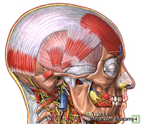
temporalis fascia
Temporalis is covered by a thick fibrous fascia, into which some of the superficial fibres insert. This creates a connection with the epicranium, the fascia covering the head and therefore the small upper neck muscles and frontal muscles. This is one reason why, if the muscle is tight or the jaw action dysfunctional, there can be issues with headaches and it can affect other structures.
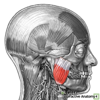
masseter
Masseter is a bit simpler to understand, though it has its moments...! Most obviously it is also important for closing the jaw or elevating the mandible. Its attachments along the bottom of the angle and ramus of the mandible, up and forwards to the zygomatic arch and zygoma let you clearly imagine how an open mouth will be closed by this muscle contracting. It is said this is proportionally the strongest muscle in the body.
When the mouth is closed and the same diagonally and upward oriented fibres contract they will draw the mandible forward, protracting it.
Now, we can't quite leave it there as this muscle also has a deep part, a section that attaches a little further back on the zygomatic arch and runs parallel to the back of the body of the mandible to the middle of the body and the lower coronoid process. Imagine that the jaw has been protracted by those superficial fibres and the mandible is further forward; the deep, more posterior ones are now angled diagonally and so if they contact will draw the mandible back again.
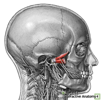
lateral pterygoid
The lateral pterygoid is attached to the disc, capsule and the head and neck of the mandible. From here it runs inwards to parts of the sphenoid bone. Most of the fibres are horizontal, thus perfectly positioned to protract the mandible and help that disc and condyle on its trip down the articular eminence. Since the fibres are medial to the mandible, when only one side contracts it can also assist in the side to side movements of the jaw.

med ptery
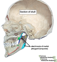
Med Ptery Atts
The final one of the four is the medial pterygoid. Like the lateral it is also on the inside of the joint and runs inwardly parallel to the masseter. Attaching from the inside of the angle of the mandible, it runs forward and up diagonally to the sphenoid bone (onto which it attaches more medially than the lateral pterygoid, hence their names). With fibres at that similar oblique angle it also closes the jaw, protracts and when one side contracts, creates the side to side grinding movements so important for good mastication.
Phew. I hope that wasn't too much to take in - the pictures are really important, so if you feel so inclined please do grab an anatomy book or find other images to help you really get to grips with them. Better still, if you can get your hands on a skull you will be able to get the 3-d locations clearer.
Remember too, one of the basics of many bodywork therapies: the body is all connected. The fascial links, functional pulls from other muscles another, issues with neural or vascular supply, what happens when the body is in different positions - e.g. in this case, the head back vs the head forward, affects the opening of the jaw... all affect the function of other parts.
I first got interested in the TMJ when someone said years ago that the positioning of the feet and a person's gait could reflect and affect all the way up into the jaw. This has two implications: when we work on any area we may initiate far reaching effects and, if we are presented with a client with a TMJ issue, we should think about its anatomical structure and function, but remembering to widen our awareness for clues in other parts of the body too.
Acknowledgement Citation
Images by Adam Interactive Anatomy
Comments:
-
No Article Comments available