Positive Health Online
Your Country

![[Image: https://www.water-for-health.co.uk/]](/img/original/BannerAvatar/4046.12831f50177f8848ee87b8e94411d6b3.jpg "https://www.water-for-health.co.uk/")
![[Image: http://www.abundanceandhealth.co.uk]](/img/original/BannerAvatar/2836.df7d2315253110ff5b3a566f94871f99.jpg "http://www.abundanceandhealth.co.uk")
![[Image: https://peacegifts.shop/]](/img/original/BannerAvatar/3906.e1d1ebc07ce7530bbb3d6cf426966b6f.jpg "https://peacegifts.shop/")
![[Image: http://www.balens.co.uk]](/img/original/BannerAvatar/2148.50367fa908013fae0a7f1a171543ef92.jpg "http://www.balens.co.uk")
![[Image: http://www.drsgoodman.com/books-goodman/52-nutrition-and-cancer]](/img/original/BannerAvatar/1859.1296090fd8385e7ceff51c8011559097.jpg "http://www.drsgoodman.com/books-goodman/52-nutrition-and-cancer")
![[Image: Turning Point]](/img/original/TopicbannerAvatar/981.28945ea95f14bc185effb4ae6d8a75ab.jpg "Turning Point")
![[Image: ISNS International Science and Nutrition]](/img/original/TopicbannerAvatar/4310.3fe1bd582f0b1b36c39fca00e3464581.jpg "ISNS International Science and Nutrition")
The Importance of Blood Flow and Evening Primrose Oil in ME
by Leslie Owen Simpson(more info)
listed in cfs me long covid, originally published in issue 169 - April 2010
Introduction
The study of blood is shared between two branches of medical science, namely haematology and haemorheology (blood rheology). While haematology deals with the cellular and chemical make up of blood, haemorheology relates to those physical factors of the blood which influence the rate of blood flow, such as plasma viscosity, whole blood viscosity and red cell deformability. For unexplained reasons, blood rheology is not taught at medical schools, and medical students will not be made aware of the thixotropic nature of blood, i.e. that there is an inverse relationship between the rate of blood flow and blood viscosity. Thus the slower the rate of blood flow, the higher is the viscosity of the blood. As a result, general practitioners will not be aware of the clinical implications of altered blood rheology except for a small number of disorders in which increased blood viscosity is an accepted feature. Polycythemia rubra vera is a condition in which the number of red cells is increased greatly, and this is manifested as increased blood viscosity with a variety of dysfunctional features. But when the excess of red cells is removed, normal tissue function is restored. Myelomatosis is a condition in which the synthesis of myeloma protein increases blood viscosity. When the myeloma protein is removed by the use of plasmapheresis, normal rates of blood flow are restored.
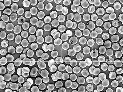
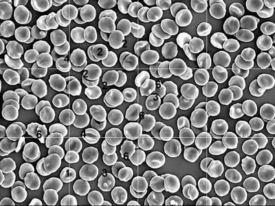
Examples of the red cells in two cases of immediately fixed blood samples from people with chronic ME. Chronic ME 1 is an example with high values for flat cells, while Chronic ME 2 shows high values for cells with altered margins in addition to increased flat cells.
Chronic ME 21. Biconcave discocytes. According to textbooks all red cells have this form.
2. Flat cells.
3. Cells with surface changes such as bumps or ridges.
4. Early cup forms. (stomatocytes) These cells are prominent in acute ME.
5. There are no examples of late cup forms which are swollen and dimpled cells.
6. Cells with altered margins. (echinocytes)
It is important to recognize that the reduced rate of blood flow occurs when blood rheology is altered, for example by increased plasma viscosity due to increased lipids, immunoglobulins or the stress hormones, or by a reduction in red cell deformability; it is possible to address such factors and to restore the rate of blood flow to normal. Therefore, the fact that the dysfunctional states associated with increased blood viscosity are reversible when the blood viscosity is restored to normal levels, is of some significance from the patient's perspective. And it is the recognition of such changes that is essential to obtain an understanding of the pathophysiology of ME, and how the condition might be managed most effectively.
ME – An Historical Perspective
For anyone interested in ME, Dr Melvin Ramsay's book Myalgic encephalomyelitis and post-viral fatigue states, second edition[1] is essential reading. Ramsay emphasized that he had never accepted the term 'Postviral fatigue states'. After a TV programme on the subject of myalgic encephalomyelitis, in which an immunologist had stated, "ME and PVFS are regarded as synonymous," Ramsay wrote, "I realized that my objection to the latter term was fully justified and that it was incumbent on me to show that the statement was blatantly untrue. It is fortunate that a second edition of my monograph affords me an opportunity to demonstrate that the clinical features of myalgic encephalomyelitis provide a sharp contrast to all forms of postviral fatigue syndromes."
An intriguing feature of Dr. Ramsay's views on the clinical features of ME was his recognition of the muscle phenomena, cerebral dysfunction and circulatory impairment in the disorder. Even though he recognized the cold hands and feet as evidence of impaired peripheral blood flow, he made no suggestion that the muscle and central nervous system dysfunction might be a consequence of the circulatory impairment. However he considered that ME had three distinctive features.
- ...A unique form of muscle fatigability whereby even after a minor degree of physical effort, three, four, or five days or longer, elapse before full muscle power is restored;
- ...Variability and fluctuations of both symptoms and physical findings in the course of a day;
- ...An alarming tendency to become chronic. In addition, he noted that in many chronic cases, remissions and relapses persisted for many years. "The fact that remissions occur has important implications for the pathophysiology of ME as such returns to normal health, no matter how briefly, could not occur if the causal factor was a persistent viral infection, as has been claimed."
The New Zealand Experience
In the early 1980s, when I was exploring the effects of filtering EDTA-anticoagulated blood through Millipore filters with 5 micron pores at different negative filtration pressures, it was found that blood samples from blood donor samples provided reproducible results. About that time ME had become a recognizable problem and had become a clinical interest of Professor Campbell Murdoch, head of the Department of General Practice. Through his good offices, blood samples were obtained from 22 women and 11 men who were suspected of having ME. The blood filtration times for the ME samples were longer that those from blood donors, and blood samples taken from those who were severely unwell at the time of sampling had the longest filtration times.[2] As people with multiple sclerosis (MS) also suffer from tiredness, a similar study revealed that MS blood was poorly filterable also. In addition, increased blood viscosity was reported.[3]
Those findings gave rise to the thought that scanning electron microscopy of the red blood cells might provide information about the observed reduction in blood filterability. By sheer chance, it was decided to adapt a technique used for the study of theatre samples, which involved immediate fixation of the blood sample. This involved the addition of 3 to 5 drops of blood to 5ml of a 'fixative', a special solution which prevented the red cells from changing shape. Assessment of the immediately fixed red cells showed very quickly that the currently accepted view that all red cells were doughnut shaped, biconcave discocytes, was not sustainable. However, the accepted status involves red cells which had been exposed to an anticoagulant and washed in physiological saline prior to fixation, and it is the treatment which transforms all red cells to biconcave discocytes. But the effects of fixation had been noted earlier, as in 1976, Miller et al [4] had reported that immediately fixed red cells from muscular dystrophy patients were different from those which had been taken into an anticoagulant and washed prior to fixation.
Immediately fixed blood samples from blood donors had red cells which could be classified on the basis of simple observable features, into six different shape classes. In 1989, the British Journal of Haematology published the first report of these findings as a 'rapid paper'.[5] But 20 years later, students are still taught that all red cells are biconcave discocytes. This unwillingness to recognize the features of red cells are shown clearly in a two-volume reference work An atlas of blood cells.[6] "Figure 12a shows 33 stained red cells and the caption draws attention to a normal erythrocyte which is arrowed and shows a clear centre ...due to the biconcavity of the cell." However, only one of the 33 cells shows this feature and there was no comment about the 32 cells which lacked clear centres. Figure 12b shows 39 cells with the caption stating, "...erythrocytes appear as biconcave discs by scanning electron microscopy," but only six or seven cells show that form. There are clearly discernible flat cells, cells with ridges and dimpled cells, none of which excited comment.
To some extent these conflicting viewpoints concerning red cell shape, draw attention to the fact that the red cell loses its nucleus as it leaves the bone marrow. It is possible that the enucleate nature of the cell renders it highly susceptible to changes in the internal environment. The study of blood samples from healthy subjects after episodes of physical and mental stress, and from patients with a variety of chronic disorders showed changed red cell shape populations.[7]
Scanning electron microscopy of blood samples from 102 ME people revealed lower than usual numbers of biconcave discocytes and higher numbers of cup forms (stomatocytes).[8] Similar results were obtained from an additional 60 females and 39 males who suffered from tiredness and easy fatigability, and the results were presented at the Cambridge Symposium on ME in 1990.[9] Although the majority of samples showed high percentages of cup-transformed red cells, there were small numbers of cases with high values for flat cells or cells with altered margins. During the next two years the latter changes became dominant, leading to the proposal that high values for cup forms was a marker for acute ME, while increased flat cells or cells with altered margins were indicators of chronic ME. This shift from acute to chronic status was well shown in a report of the red cell shape populations of 1558 female and 620 male members of ME organisations in New Zealand, Australia, South Africa and the UK.[10] In both sexes, high values for flat cells was the dominant change, and about 15% had no abnormal values, which suggested that they might have been in remission at the time of sampling.
An interesting observation was that in 1977, increased stomatocytes (cup forms) had been reported in patients with Huntington's Disease.[11] Subsequently it was reported that Huntington's patients had reduced cerebral blood flow which correlated with cognitive functioning [12], but there was no comment about the implications for cerebral blood flow of the red cell changes noted in the 1977 report.
Because changed shape populations of red cells can be expected to impair cerebral blood flow, it is not surprising that in 1983 Swank et al should find impaired cerebral blood flow in MS patients.[13] They noted, "The decreases in CBF were accompanied by parallel increases in disability which were so clearly linked temporally that a cause and effect relationship seems possible." Dr DC Costa presented his findings from a SPET study in the brainstem of ME patients at the annual general meeting of the British Nuclear Medicine Society on March 30, 1994. He reported that brain blood flow in ME patients was significantly lower than in healthy subjects. But the findings were reported by Costa et al in 1995,[14] and amongst other things noted that ME/CFS patients, "...had a generalized reduction in brain perfusion." Thus, in three different disorders in which changed red cell shape populations have been demonstrated, impaired cerebral blood flow is a common feature.
Capillary Blood Flow
It should be emphasized that changes in the physical properties of the blood are only one factor in determining the rate of blood flow in the microcirculation. As the average diameter of capillaries is about 3.5 to 4 microns, this means that most capillaries will be smaller than the 8 micron diameter of red cells. This situation draws attention to the importance of red cell deformability in blood flow in the microcirculation. While healthy subjects may show changed red cell shape populations after episodes of mental or physical stress, they do not develop the health problems of ME people. This implies that in some way those who develop ME are different. In 1992 it was suggested that, "...subjects with the symptoms of tiredness and high percentages of nondiscocytic red cells in their blood would have smaller-than-usual capillaries, i.e. those with mean capillary diameter falling in the first quartile of a size distribution. Subjects with this characteristic would always be at risk of red cell shape impairment of capillary blood flow."[15] Given the widely variable distribution of symptoms in ME people, it is envisaged that clusters of small capillaries would be distributed randomly throughout the body. As it is not possible to increase the size of a capillary, this means that treatments should be aimed at increasing red cell deformability.
Remissions
According to Ramsay[1] chronic ME may take two forms. In one form there is, "...a recurring cycle of remission and relapse." But there are no remissions in the second form. He reported the cases of three doctors who had contracted ME between 1955 and 1958 and who were still having remissions and relapses thirty years later. Rather surprisingly, the mechanism(s) of remissions remain uninvestigated, and they appear to be ignored by most investigators. This is well shown by the situation in the book by Hyde et al [9] where of the many authors, only two mentioned remissions. Dr Snow wrote on p105, "The disease over the years has demonstrated its remitting and relapsing nature." On p530, Dr Loblay stated, "...the fluctuating course, both short-term and long-term, the occurrence of spontaneous remissions (occasionally full recovery) even after prolonged illness." But neither author commented on the implications of remissions.
In the request form for red cell shape analysis, there is a section which requests a statement about wellbeing. Patients are requested to indicate if they are well with no symptoms, well with slight symptoms, slightly unwell, moderately unwell or severely unwell. About 9am a young woman brought a blood sample to the office, and it was noted that she was well with no symptoms. It was pointed out that if she was well, then it was likely that the result from the sample would be normal. Late in the afternoon of the same day she brought in another sample, taken when she was severely unwell. She reported that for an unexplained reason she had 'crashed' about 4pm. The first sample showed normal values while the second sample was grossly abnormal. As this was the first observation that remissions were associated with normal blood values, the finding stimulated a search for more information.
A panel of ME volunteers (37 females, 11 males) who had been diagnosed as having ME by a physician at least two years previously, agreed to meet every four weeks for forty weeks. At each meeting a blood sample was obtained and their state of health was recorded in terms of symptoms and their perceived level of wellbeing. The most striking feature of the results was the range of variability of their health problems. At one extreme, five women had abnormal values in all eleven blood samples, which were associated with severe symptoms and poor wellbeing. While it is possible that the women may have experienced remissions in the four-week interval, it is also possible that they had the non-remitting form of ME. At the other extreme was a woman who had six of her eleven samples showing normal values. When the results from both sexes were combined, the median result was to have two normal blood tests with normal wellbeing during the forty weeks of the study. On the basis of these findings, it is reasonable to conclude that remissions are not uncommon events in people with ME.
Even though the mechanism of remissions remains unexplained, they appear to be an important part of the pathophysiology of ME. Just what factor (or factors) can switch 'off' to restore normal levels of wellbeing and red cell shape populations remains unknown. While it is recognized that episodes of emotional or physical stress can initiate a relapse, it is not known if such events are necessary for the relapsing mechanism to be switched 'on'. But if the changes which lead to remissions were understood, then maybe the management of ME could be improved.
The Treatment of ME
Poor blood filterability in ME patients can be taken as an estimate of reduced red cell deformability, which together with changed shape populations of red cells, will impair capillary blood flow. Therefore, as both factors are normalized during remissions, the major target of treatment should be to attempt to restore red cell deformability. Although there are several agents which could be effective, there are no random controlled trials which could assess their effectiveness. The relatively rapid adoption of terms such as ME/CFS and CFS/ME by government officials makes it very unlikely that sufficient ME people could be identified for a random controlled trial. And with the general acceptance of CFS as a correct diagnosis, it would be very difficult to obtain financial support for such trials. There are several agents which have been shown to improve red cell deformability in different conditions as well as in ME.
1. Acute ME: Vitamin B12, hydroxocobalamin. (Neo-cytamen)
Although hydroxocobalamin is recommended for the treatment of pernicious anaemia, Ellis and Nasser[16] found its greatest use was in the treatment of tiredness. In 1989 a patient came to request a blood test because she felt so well after receiving an injection of hydroxocobalamin. The blood test revealed that her improved wellbeing was associated with a marked reduction in cup-transformed red cells. Injections of the B12 at about 12 day intervals resulted in the maintenance of her improved health. Subsequent pre- and post-B12 blood samples provided by general practitioners showed that only 50% of cases responded, and this situation remains unexplained.
2. Chronic ME
Evening Primrose Oil
The most important component of evening primrose oil is the gammalinolenic acid which is eventually converted to prostaglandin E1 (PGE1); the objective of taking the oil is to raise the blood levels of PGE1. Manku et al[16] reported that 4 grams daily of Efamol produced a significant increase in the blood levels of PGE1. Kury et al[17] used a spin-label technique to show that PGE1 increased the fluidity of the lipid bilayer of the red cell membrane. The resulting increase in red cell deformability, was shown by Rasmussen et al[18] as an improvement in blood filterability. On the basis of these findings, it has been suggested that people suffering from chronic ME should take 4 grams of evening primrose oil daily, and many have reported how the treatment had changed their lives. However, not all patients benefited and it was not possible to identify which patients would not respond. But the lack of a response could indicate the use of a spurious oil. In 1980, the New Zealand Institute of Chemistry assessed eleven brands of evening primrose oil and found that only two of the eleven were authentic. In 1997 we were able to confirm that Efamol, Naudicelle and EPO were authentic evening primrose oils, but this does not exclude the possibility that there are other authentic brands.
It was recommended that when assessing the effects of an oil supplement, this should be done after six weeks on the oil. Unless a clear benefit was discernible, the supplement should be stopped and another assessed.
Fish oil
There is much published evidence that a diet rich in oily fish greatly reduces the incidence of heart disease, although in many reports the beneficial effects on blood flow are not recognized. Kamada et al[20] used a spin-label technique to show that the omega-3 fatty acids in sardine oil increased the fluidity of the lipid bilayer of the red cell membrane. This is the same change that Kury et al [17] had obtained with PGE1. At least 6 grams daily of fish oil are needed to obtain a sustained benefit.
Other Agents
There is evidence that the beneficial effects of extracts of Gingko biloba could be helpful in ME people. The drug pentoxifylline might also be useful. A Danish man with the typical changes of ME got no benefits from six weeks of evening primrose oil or from 6 weeks of fish oil. However he responded to a daily intake of 1200mg of pentoxifylline.
Conclusions
- There is no doubt that the poor filterability of ME blood and the abnormal populations of red cell shapes indicate that capillary blood flow will be impaired in ME. If, as suggested, ME people have smaller than usual capillaries this would provide an anatomical basis for the disorder, but the potentially treatable aspect of the problem is the reduced red cell deformability.
- The significance of the capillary blood flow problem is highlighted during remissions, when normal tissue function is restored and the abnormal features of the blood disappear.
- Taking sufficient evening primrose oil (at least 4 grams daily) to provide a significant rise in the blood levels of prostaglandin E1 provides effective relief by increasing red cell deformability. However no controlled trials have been carried out to assess the benefits of the treatment in chronic ME.
- In acute ME, vitamin B12 as hydroxocobalamin provides relief in only 50% of cases, and the reason for this remains unexplained.

References
1. Ramsay AM. Myalgic encephalomyelitis and postviral fatigue states. Second edition. London, Gower Medical Publishing for the ME Association. 1988.
2. Simpson LO, Shand BI, Olds RJ. Blood rheology and myalgic encephalomyelitis: a pilot study. Pathology 18:190-192. 1986.
3. Simpson LO, Shand BI, Olds RJ, Larking PW, Arnott MJ. Red cell and haemorheological changes in multiple sclerosis. Pathology 19:51-55. 1987.
4. Miller SE, Roses AD, Appel SH. Scanning electron microscope studies in muscular dystrophy. Arch Neurol 33: 172-174. 1976.
5. Simpson LO. Blood from healthy animals and humans contains nondiscocytic erythrocytes. Br J Haematol 73: 561-564. 1989.
6. Zucker-Franklin D, Greaves MF, Grossi CE, Marmont AM. An atlas of blood cells, vol 1. Philadelphia, Lea and Febiger, p52. 1981.
7. Simpson LO. Red cell shape in health and disease. In: Swamy NCV, Megha Singh (eds) Physiological Fluid Dynamics III, Narosa Publishing House, New Delhi, 230-235. 1992.
8. Simpson LO. Nondiscocytic erythrocytes in myalgic encephalomyelitis. NZ Med J 102: 106-107. 1989.
9. Simpson LO. The role of nondiscocytic erythrocytes in the pathogenesis of myalgic encephalomyelitis. In: Hyde BM, Goldstein J, Levine P (eds). The clinical and scientific basis of myalgic encephalomyelitis/ chronic fatigue syndrome. The Nightingale Research Foundation, Ottawa, p597-605. 1992.
10. Simpson LO, Herbison GP. The results from red cell shape analyses of blood samples from members of myalgic encephalomyelitis organisations in four countries. J Orthomol Med 12: 221-226. 1997.
11. Markesbery WR, Butterfield DA. A scanning electron microscope study of erythrocytes in Huntington's Disease. Biochim Biophys Res Commun 78: 560-564. 1977.
12. Tanahashi N, Meyer JS, Ishikawa Y, Kandula P, Mortel R, Rogers RL Cerebral blood flow and cognitive testing correlate in Huntington's Disease. Arch Neurol 42: 1169-1175. 1985.
13. Swank RL, Roth JG, Woody DC. Cerebral blood flow and red cell delivery in normal subjects and in multiple sclerosis. Neurol Res 5: 37-54. 1983.
14. Costa DC, Tannock C, Brostoff J. Brain stem perfusion is impaired in chronic fatigue syndrome. QJM 88: 767-773. 1995.
15. Simpson LO. Chronic tiredness and idiopathic chronic fatigue – a connection. N Jersey Med 89: 211-218. 1992.
16. Ellis FR, Nasser S. A pilot study of vitamin B12 in the treatment of tiredness. Br J Nutr 30: 277-283. 1973.
17. Manku MS, Horrobin DF, Morse N, Kyle V, Jenkin K. Reduced levels of prostaglandin precursors in the blood of atopic patients: defective delta-6-desaturase as a biochemical basis for atopy. Prost Leuko Med 9: 615-628. 1982.
18. Kury PG, Ramwell PW, McConnell HM. The effect of prostaglandin E1 and E2 on
the human erythrocyte as monitored by spin labels. Biochim Biophys Res Commun 56: 478-483. 1974.
19. Rasmussen H, Lake W, Allen JE. The effects of catecholamines and prostaglandins upon human and rat erythrocytes. Biochim Biophys Acta 411: 63-73. 1975.
20. Kamada T, Yamashita T Baba Y. Dietary sardine oil increases erythrocyte membrane fluidity in diabetic patients. Diabetes 35: 604-611. 1986.
Comments:
-
No Article Comments available