Positive Health Online
Your Country

![[Image: https://www.water-for-health.co.uk/]](/img/original/BannerAvatar/4046.12831f50177f8848ee87b8e94411d6b3.jpg "https://www.water-for-health.co.uk/")
![[Image: http://www.abundanceandhealth.co.uk]](/img/original/BannerAvatar/2836.df7d2315253110ff5b3a566f94871f99.jpg "http://www.abundanceandhealth.co.uk")
![[Image: https://peacegifts.shop/]](/img/original/BannerAvatar/3906.e1d1ebc07ce7530bbb3d6cf426966b6f.jpg "https://peacegifts.shop/")
![[Image: http://www.balens.co.uk]](/img/original/BannerAvatar/2148.50367fa908013fae0a7f1a171543ef92.jpg "http://www.balens.co.uk")
![[Image: http://www.drsgoodman.com/books-goodman/52-nutrition-and-cancer]](/img/original/BannerAvatar/1859.1296090fd8385e7ceff51c8011559097.jpg "http://www.drsgoodman.com/books-goodman/52-nutrition-and-cancer")
![[Image: ICAN International Clinical Aromatherapy Network]](/img/original/TopicbannerAvatar/4442.566a83a07de79de73b9c738f7fb6458a.jpg "ICAN International Clinical Aromatherapy Network")
Case Study Issue 122: Successful Use of PDT for Vascular Tumour
listed in case studies, originally published in issue 122 - April 2006
Vascular lesions in children are more common than we might think. Whether it be birthmarks or more sinister haemangiomas in the limbs, the heart and lungs, as well as the eyes, face and head, their figures are probably widely un-reported as they fall under so many different headings.
There are a number of established treatments, but now a newcomer to the list looks really promising – Photodynamic Therapy (PDT). Last December, physicians at the National Medical Laser Centre (NMLC) at London's University College Hospital conducted the MRI and MRA scans on the vascular tumour that has blighted the first 21 years of Louise Longman's life. There was barely a trace of the tumour eight weeks after the PDT.
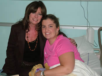
Pre-op-Louise Longman
Louise is my daughter who has suffered terribly over the years. Her story began when she had surgery in Nottingham's Queen's Medical Centre when she was six. The tumour was in her left forearm, and she was left with severe muscle and tendon loss. The lesion re-grew as she entered her teens, and she would often cry out if her arm was even brushed against. Her mood swings were understandably quite violent as she struggled to cope emotionally and physically.
Many treatment options were tried over the years, with percutaneous sclerotherapy the last in the line of serious options which didn't produce the desired results. Surgery and radiotherapy were ruled out due to the likely further destruction of tendons and muscles.
We became aware of PDT – Photodynamic Therapy – and its selective ability to destroy cancer tumours in the mouth, oesophagus and skin. Its use in the bile ducts, pancreas and lungs in the UK at the NMLC, is producing some outstanding results, while prostate cancer patients are among those also gaining great benefit.
It was Professor Stephen Bown and Colin Hopper at the NMLC who were persuaded by Louise to take on her case in July 2005. They did so, I believe, in sympathy for her plight. Some months before, Louise had completed a series of PDT treatments in Russia that was an expensive and abject failure. The group's lack of current knowledge was so pronounced, when compared to the work of the team at the NMLC.
PDT – in this case – used the Foscan drug from German company, Biolitec. It is approved for head and neck cancers, while Colin Hopper has been increasingly experimenting with Foscan and PDT for skin and facial cancers. Basal Cell Carcinoma (BCC) and squamous cancers are proving to be highly receptive to this combination therapy.
Foscan is absorbed into the tumour and is activated with light of the right wavelength [652 nm red light] – usually 96 hours later. It changes oxygen molecules into singlet oxygen that cells can't tolerate. Because no heat is used or generated, healthy cells destroyed will re-grow within the undamaged underlying scaffold of the tissue, leaving the body with little or no scar tissue and a near perfect cosmetic effect. Usually, just a single PDT treatment is required, while a vial of Foscan (£4,400) can be split between a number of patients treated simultaneously, depending upon the drug volumes required.
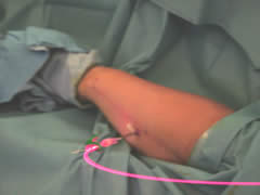 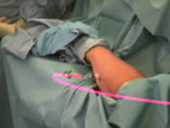 Hollow fibres were placed into the arm down which the laser was directed. |
One Friday afternoon we headed for the NMLC to have the drug injected. From that moment on, Louise's body became light sensitive. She needed to wear dark clothing and dark glasses, protecting herself from possible skin burn reactions.
The following Tuesday she returned for the light treatment. Hollow fibres were placed into the arm down which the laser was directed, and just 20-seconds of light exposure was needed to trigger the reaction in the target area.
Just 24-hours later Louise was already noting a series of dramatic changes to the tumour area. It was no longer hard and the swelling had reduced. Warnings that she would need high strength pain killers proved to be ill-founded. The response was that quick.
Given the volume of drug used, within two weeks of the Friday injection she was able to dispense totally with any light exposure precautions, having weaned herself gradually back to full exposure.
Louise had to wait eight weeks for the MRI and MRA scan, allowing for the dead cells to be taken away. The news was spectacular. The entire tumour had disintegrated along its length, breadth and depth, with some minor fatty tissue the only reminder of where it had been. She is now pain free. The doctors say it is highly unlikely that it will ever return.
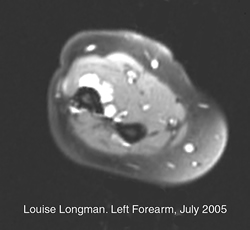
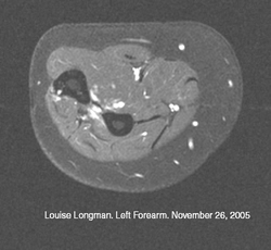
MRI scans: Before and after PDT treatment
Louise is now appealing for funding for the medical team to pay for new research and patient treatments. Says Colin Hopper: "Louise's successful treatment opens up so many other possibilities, including the treatment of other vascular lesions, including birthmarks and haemangiomas. Now we have to take the next steps forward, and advance our understanding of PDT to the next level in this vital area.
Further Information
Please email David Longman via killing.cancer@virgin.net. Donations to help further PDT research at the Centre can be made via the KILLING Cancer www.killingcancer.co.uk, or via cheques sent to KILLING Cancer at PO Box 47488, London, N13 4US.
Comments:
-
No Article Comments available